Introduction
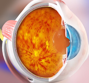
Retinal vein occlusion occurs when one of the small veins responsible for draining blood from the retina becomes blocked, usually due to a blood clot or narrowing of the blood vessels. The retina, a light-sensitive layer at the back of the eye, relies on a healthy blood supply to provide oxygen and nutrients to nerve cells that convert images into signals for the brain. When blood flow is obstructed, swelling and damage can occur, leading to vision problems. This condition most commonly affects people over 60 and is often associated with underlying health issues such as high blood pressure, diabetes, and high cholesterol. Early diagnosis and management are vital to prevent serious complications like macular oedema and neovascular glaucoma.
Causes and Risk Factors of Retinal Vein Occlusion
What Causes Retinal Vein Occlusion?
Retinal vein occlusion happens when the veins that carry blood away from the retina become blocked, disrupting normal blood flow and leading to swelling and damage in the eye. Understanding the causes and risk factors helps identify those at greater risk and emphasises the need for regular eye exams.
Common causes and risk factors include:
- High Blood Pressure – A leading factor that damages blood vessels and contributes to vein blockage.
- Diabetes – Causes damage to blood vessels, increasing the risk of occlusion.
- High Cholesterol and Other Vascular Diseases – Contribute to atherosclerosis, narrowing arteries and veins.
- Narrow Retinal Veins – Some people have naturally narrower veins, increasing their susceptibility.
- Age – Mostly affects individuals over 60 years old.
Awareness of these factors is essential to control underlying health issues and reduce the risk of retinal vein occlusion and its complications.
Types of Retinal Vein Occlusion
Different Forms of Retinal Vein Occlusion
Retinal vein occlusion is classified based on the location and extent of the vein blockage. Identifying the type is important for prognosis and guiding treatment decisions.
Central Retinal Vein Occlusion (CRVO)
- Description:
Blockage occurs in the main vein draining the entire retina, causing widespread retinal swelling and vision loss. - Who it affects:
Typically affects older adults with vascular risk factors such as hypertension and diabetes. - Speed of progression:
Vision loss can be sudden and severe; complications may develop over weeks to months.
Branch Retinal Vein Occlusion (BRVO)
Description:
Blockage affects one of the smaller branches of the retinal vein, leading to localized retinal swelling and vision problems.Who it affects:
More common than CRVO, often linked to arteriosclerosis and blood vessel narrowing.Speed of progression:
Symptoms may develop gradually or suddenly depending on the extent of the blockage.
Hemispheric Retinal Vein Occlusion
- Description:
Involves blockage of either the upper or lower half of the retinal vein system, affecting a large portion but not the entire retina. - Who it affects:
Patients with intermediate severity between CRVO and BRVO. - Speed of progression:
Variable, depending on the severity and presence of complications.
Each type requires a tailored approach to monitoring and treatment to prevent vision loss and manage complications effectively.
Early Signs & Symptoms
Common Symptoms of Retinal Vein Occlusion
Recognising the early symptoms of retinal vein occlusion is important for prompt diagnosis and treatment to preserve vision and prevent complications. Symptoms may appear suddenly or worsen gradually.
- Blurred or distorted vision – Vision may become hazy or less clear, often in one eye.
- Sudden vision loss – In some cases, vision may be lost rapidly or partially in the affected eye.
- Floaters or dark spots – Patients may notice small spots or shadows drifting across their vision.
- Visual field defects – Loss of vision in part of the visual field, depending on the area affected.
- Eye discomfort or pressure – Occasionally, increased eye pressure from complications may cause discomfort.
Early detection allows for timely treatment to manage symptoms and reduce the risk of permanent vision loss.
Diagnosis and Treatment of Retinal Vein Occlusion
Retinal vein occlusion is diagnosed through a thorough eye examination that includes visual acuity testing, dilated fundoscopy to inspect the retina, and measurement of intraocular pressure. Imaging tests such as fluorescein angiography and optical coherence tomography (OCT) help evaluate the extent of retinal swelling and abnormal blood vessel growth. Blood tests may also be performed to check for underlying conditions like diabetes and high cholesterol.
While there is no treatment to reopen the blocked vein, managing the underlying causes such as high blood pressure and diabetes is essential to prevent further damage. Complications like macular oedema can be treated with focal laser photocoagulation or anti-VEGF injections to reduce swelling. Neovascular glaucoma, a serious complication, is managed with pan-retinal laser photocoagulation and sometimes anti-VEGF therapy. Early and ongoing treatment is key to preserving vision and preventing blindness.
Why Timely Diagnosis Matters
Early diagnosis of retinal vein occlusion is critical to prevent vision-threatening complications and preserve eyesight. Prompt detection allows your ophthalmologist to manage underlying health issues and intervene before irreversible damage occurs.
Timely diagnosis enables your ophthalmologist to:
- Identify retinal swelling and abnormal blood vessel growth early
- Monitor the progression and tailor treatment plans accordingly
- Prevent complications such as macular oedema and neovascular glaucoma through timely therapies
- Control underlying conditions like hypertension and diabetes to reduce further risk
- Educate patients on managing systemic health for long-term eye health
Early intervention improves treatment success and helps maintain the best possible vision and quality of life.
Continue Learning About Other Eye Conditions
Other Eye Conditions
The eyes are the most complex sensory organ in our bodies. The eyes provide vision by recording images of our surroundings that the brain will interpret. Although the eye measures only about an inch...
Blepharitis
Blepharitis is a common condition characterised by inflammation of the eyelids, typically affecting both eyes along the edges of the eyelids. It causes redness, swelling, itchiness, and sometimes crusting...
Worried About Your Vision?
Schedule a consultation with Mr. Mo Majid to evaluate your eye health.
Quick Answers About Retinal Vein Occlusion
What causes retinal vein occlusion?
It occurs when a blood clot blocks the veins draining blood from the retina, often linked to high blood pressure, diabetes, or vascular disease.
Can retinal vein occlusion cause sudden vision loss?
Yes, vision loss can be sudden or gradual, depending on the extent and location of the vein blockage.
Who is most at risk of retinal vein occlusion?
People over 60 with underlying conditions like hypertension, diabetes, and high cholesterol are at higher risk.
Is there a cure for retinal vein occlusion?
While the blocked vein cannot be reopened, treatments exist to manage complications and control underlying diseases to prevent further vision loss.


