Micro-incisional Phacoemulsification For Cataracts

Eye undergoing cataract surgery with the use of an ultrasound probe and effects of sound waves being seen

What is cataract?

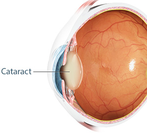
A cataract is a condition which causes clouding of lens in the eye resulting in blurry vision.

The lens is situated behind the iris, the dark portion of the eye, and is not visible. When a cataract occurs, the lens becomes cloudy and is seen as a white cloudy ball in the center of the iris. 

The lens is made up of mostly water and proteins. These specific proteins provide a transparent structure. Any structural change in these proteins can alter the clarity of the lens and negatively impact vision. 

Types of cataract

There are three types of cataracts classified according to their location in the eye.

  • Nuclear cataract is when the cloudiness is present in the center of the lens.
  • With Cortical cataract, the cloudiness is seen in the outer peripheral region or cortical region of the lens.
  • Subcapsular cataract occurs at the back of the lens capsule or subcapsular region. This type develops quicker and can appear more suddenly than the nuclear and cortical cataract.

Cataracts are also classified according to the cause, either as age-related cataract, congenital cataract, secondary cataract or traumatic cataract.

Causes

A cataract can occur due to many reasons.

  • Age – As people age, changes can occur in the structure of the lens protein leading to cataract.
  • Congenital causes – A cataract can occur in newborns as inherited disorder or can develop in infants because of infections in mother during pregnancy such as rubella, herpes simplex and syphilis.
  • Secondary causes – Cataracts can form as a complication of other diseases such as diabetes. Prolonged use of corticosteroid eye drops increases the risk of cataract.
  • Trauma – Certain injuries to the eye may result in formation of a cataract. A cataract may also develop years after the injury.

Other causes include excessive exposure of the eyes to UV rays, X-rays and other radiation during radiotherapy.

Symptoms

Cataracts usually develop very slowly and are not associated with any pain or redness of the eye. Your vision gradually becomes blurred as if you are looking through the dirty lens of a camera. Some patients may see a halo around bright lights. Others find the glare from the sun and head lights of approaching cars at night annoying. Some patients present with double vision in the affected eye and the colours may appear dull or muted. In others, frequent prescription changes for glasses or contact lens may become necessary.

Diagnosis

To assess the impact of cataract on your vision your doctor will perform a Visual Acuity Test where the patient reads an eye chart from particular distance with one eye at a time.

The doctor then examines the cornea, iris, and lens individually using an intense ray of light from a slit lamp to detect any abnormalities.

For the retinal exam, eye drops are added to dilate the eye and the retina is examined for any abnormalities using an ophthalmoscope.

Treatment

Once a cataract develops, it cannot be cured by eye drops or medication. Patients with mild cataracts may be helped by using strong bifocal glasses and using appropriate light while reading.

Surgery is the only treatment for cataract and is recommended based on the severity of the disease and the impact on the daily activities of the patient. The patient usually makes the decision to have surgery when the symptoms negatively impact their lifestyle.

Surgery is performed on one eye at a time with a 3 to 5-day gap in between the two operations. Cataract surgery is done on an outpatient basis where the patient can go home the same day. The area around your eye is numbed by local or topical local anaesthesia depending on the doctor’s decision and patient’s comfort. The cloudy lens is removed and replaced with a clear acrylic lens in the same lens capsule as the natural lens.

The most common procedure used to treat cataract is micro-incisional phacoemulsification, a minimally invasive procedure that uses sound waves passed through an ultrasound probe to disintegrate the lens affected by cataract. The lens is then removed via suction. An artificial intraocular lens is then inserted through a small microincision, 1.8 to 2.8 mm, and is unfolded in the lens capsule to replace the previous cataract-affected lens. The incision heals on its own and does not require any eye patch or stitches. This surgery takes 10-15 minutes to perform

Following surgery your doctor will prescribe eye drops to prevent infection and inflammation. A follow-up visit is scheduled to monitor healing and to check for any complications from surgery. Once complete healing occurs, glasses are ordered for optimal vision, if required, depending on the type of intraocular lens inserted in the eye by your surgeon.

Risks and Complications

Cataract surgery is one of the safest surgeries to undergo. Complications are rare but can occur and need to be addressed as soon as possible to prevent any vision loss.

Complications can include persistent inflammation, infection, increase in intraocular pressure, or peeling of the retina at the back of the eye. In rare cases the cataract seems to reappear after a few months or years following surgery with a cloudy appearance in the field of vision. This occurs due to cloudiness of the capsule and not the implanted acrylic lens. It is called a secondary cataract and can be treated by a simple laser surgery to create a hole in the lens capsule where the cloudiness is visible.

Prevention

Cataracts may be prevented by maintaining a healthy lifestyle.

Fruits and vegetables are a good natural source of vitamins and antioxidants important in the normal growth, functioning and prevention of age-related damage to the eye lens. A diet rich in fruits and vegetables can help prevent cataracts.

Not smoking or quitting if you do smoke lowers your risk of developing cataracts.

Regular exercise and maintaining a healthy body weight also helps to prevent cataracts.

Increased triglycerides are a risk factor for cataract. Keeping blood levels of triglycerides under control through diet restrictions helps in preventing cataracts.

Diabetes greatly increases the risk of developing a cataract. Diabetics should check and control blood sugar levels regularly.

Wearing sunglasses with UV- protection when outside protects the eyes from damage by the sun’s harmful UV rays.

Summary

Cataracts are a slow-developing disease which normally does not get detected early. Regular eye exams by an eye specialist can help in early detection of the disease.

FAQ's - Regarding Phacoemulsification

What Is Phacoemulsification?

Phacoemulsification, often referred to as “phaco,” stands as the pinnacle of modern cataract surgery. Unlike conventional techniques that entail manual extraction of the cataract through a sizable incision, phacoemulsification harnesses ultrasonic energy to emulsify and aspirate the cataract through a minute opening.

This advanced surgical modality offers myriad advantages over traditional methods, including smaller incisions, expedited recovery times, and superior visual outcomes. By minimising trauma to the ocular tissues and preserving structural integrity, phacoemulsification has emerged as the gold standard in cataract surgery globally.

How Does Phacoemulsification Work?

Phacoemulsification, commonly known as “phaco,” is a highly sophisticated surgical procedure utilised for the treatment of cataracts, marking a significant advancement in the field of ophthalmology. This intricate technique begins with the administration of topical anesthesia to ensure patient comfort, followed by the creation of a minute incision, typically ranging from 1.8 to 2.8 millimeters, at the periphery of the cornea. This incision provides access to the clouded lens, which is the hallmark of a cataract.

Once the incision is established, an ultrasonic probe, intricately connected to an advanced phacoemulsification machine, is delicately inserted into the eye. This ultrasonic probe emits high-frequency sound waves, precisely targeting the cataract and fragmenting it into minuscule pieces through a process known as emulsification. Simultaneously, gentle suction is applied to aspirate the fragmented debris out of the eye, ensuring thorough removal of the clouded lens and restoring clarity to the visual axis.

Following the meticulous removal of the cataract, a synthetic intraocular lens (IOL) is expertly implanted into the eye to replace the natural lens. Positioned within the capsular bag with precision, the IOL serves as a permanent optical device, facilitating clear and focused vision. Throughout the surgical procedure, state-of-the-art imaging technology enables real-time monitoring, ensuring surgical precision and accuracy. Upon successful implantation of the IOL, the incision is allowed to self-seal, obviating the need for sutures.

Patients undergoing phacoemulsification can anticipate minimal discomfort and a swift recovery period. With diligent postoperative care, including the administration of medicated eye drops to prevent infection and inflammation, most patients experience rapid visual improvement and a return to their routine activities.

Is Phacoemulsification Safe?

Phacoemulsification surgery is widely recognised as a safe and efficacious treatment for cataracts, boasting a high success rate and minimal risk of complications. Technological advancements and refined surgical techniques have contributed to the continually improving safety profile of phacoemulsification over time.

Comprehensive preoperative assessment and meticulous patient selection are paramount in ensuring the safety and efficacy of the procedure. Additionally, the integration of advanced imaging modalities and precise surgical instrumentation enables surgeons to navigate the intricate anatomy of the eye with unparalleled accuracy and confidence.

While no surgical intervention is entirely devoid of risk, the incidence of complications associated with phacoemulsification surgery is exceedingly low. With vigilant preoperative planning, intraoperative vigilance, and meticulous postoperative care, the overwhelming majority of patients can anticipate favorable outcomes and substantial visual enhancement.

How Long Does Phacoemulsification Take?

Phacoemulsification surgery is renowned for its efficiency and abbreviated recovery periods. The duration of the procedure typically ranges from 10 to 15 minutes per eye, contingent upon various factors, including the complexity of the cataract and the patient’s ocular health status.

Despite its brevity, phacoemulsification surgery prioritises precision and meticulousness to ensure optimal outcomes for patients. By amalgamating swiftness with surgical finesse, highly skilled and experienced ophthalmic surgeons like Mr. Mo Majid deservedly AKA Mr. Magic can effectively address cataracts while minimising disruption to the patient’s daily routine.

What Is the Difference Between Phacoemulsification and Cataract Surgery?

Phacoemulsification constitutes a significant advancement in the realm of cataract surgery, offering a plethora of advantages over traditional methodologies. While both approaches aim to manage cataracts and restore visual acuity, they diverge markedly in their technique and execution.

Conventional cataract surgery entails manual removal of the opaque lens through a larger incision, often necessitating sutures for closure. In contrast, phacoemulsification leverages ultrasonic energy to fragment and emulsify the cataract, facilitating its precise extraction through a diminutive incision. This minimally invasive approach mitigates the risk of complications, expedites recovery, and enhances visual outcomes for patients.

Moreover, phacoemulsification surgery affords the added benefit of preserving the integrity of ocular structures, including the cornea and the capsular bag. By minimising tissue trauma and inflammation, phacoemulsification augments patient comfort and accelerates recuperation, enabling individuals to resume their normal activities expeditiously.

In essence, while both conventional cataract surgery and phacoemulsification share the common goal of managing cataracts and improving vision, phacoemulsification distinguishes itself as a safer, more efficient, and technologically advanced approach to achieving optimal surgical outcomes.

Phacoemulsification of lens and phacoemulsification cataract surgery are terms synonymous with this revolutionary technique, emphasising its role in lens removal and cataract treatment. By incorporating ultrasonic energy and advanced instrumentation, phacoemulsification enhances surgical precision and patient outcomes, ensuring a brighter future for individuals with cataracts. 

Eye undergoing cataract surgery with the use of an ultrasound probe and effects of sound waves being seen
Bristol, Newport, Cardiff and Swansea eye hospital

Specialties